Kód: 09231346
Brain Imaging
Autor Paul C. Lebby
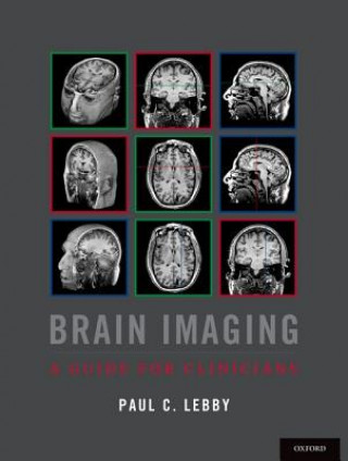
Brain Imaging: A Guide for Clinicians is designed to provide a foundation of information necessary for those wishing to integrate brain imaging into their practice, or to those who currently review brain scans but have minimal for ... celý popis
- Jazyk:
 Angličtina
 - Vazba: Brožovaná
- Počet stran: 432
Nakladatelství: Oxford University Press Inc, 2015
- Více informací o knize

Mohlo by se vám také líbit
Darujte tuto knihu ještě dnes
- Objednejte knihu a zvolte Zaslat jako dárek.
- Obratem obdržíte darovací poukaz na knihu, který můžete ihned předat obdarovanému.
- Knihu zašleme na adresu obdarovaného, o nic se nestaráte.
Více informací o knize Brain Imaging
Nákupem získáte 547 bodů
 Anotace knihy
Brain Imaging: A Guide for Clinicians is designed to provide a foundation of information necessary for those wishing to integrate brain imaging into their practice, or to those who currently review brain scans but have minimal formal training in neuroimaging. The guide covers a range of topics important to those using brain imaging, such as the strengths and weaknesses of the many different techniques currently available, the factors that may influence the use of imaging data, common pitfalls or artifacts that may be misleading to the clinician, the most appropriate techniques to use given a specific clinical question or condition, how to interpret information presented on a brain image, and also how many pathological conditions appear on a variety of brain scanning techniques or sequences. This guide also provides detailed information regarding the identification of primary brain regions, anatomical structures, systems or pathways using both two-dimensional and three-dimensional imaging techniques. A brain atlas is included using both CT and MRI sequences to facilitate the reader's ability to identify most primary brain structures. A novel color-coded system is used throughout this guide to assist the reader in identifying slice locations and orientations. Images with green borders are displayed in the axial plane, with the slice location being shown on other orthogonal image planes by a green line. Similarly, images with a red border are displayed in the coronal plane and those with a blue border are displayed using a sagittal plane; red and blue reference lines are displayed on orthogonal slices to identify the slice location. The crosshairs formed by the color-coded reference lines optimize the reader's ability to identify primary anatomical structures or pathological markers and processes. Chapters in this book progress from a general description of the clinical use of brain images and the interpretation of scans, to more complex material involving neuroanatomy and imaging technology. Real-life examples of clinical cases are integrated into all chapters of this guide. Brain Imaging: A Guide for Clinicians features hundreds of images derived from traumatic and non-traumatic pathologies to provide the reader with examples of conditions most often seen in the clinic. PEARL-PERIL sections outline critical information for the clinician, along with many tables and charts designed to provide general information required when interpreting brain images.
 Parametry knihy
Zařazení knihy Knihy v angličtině Mathematics & science Biology, life sciences Life sciences: general issues
5467 Kč
- Plný název: Brain Imaging
- Podnázev: A Guide for Clinicians
- Autor: Paul C. Lebby
- Jazyk:
 Angličtina
 - Vazba: Brožovaná
- Počet stran: 432
- EAN: 9780190239060
- ISBN: 0190239069
- ID: 09231346
- Nakladatelství: Oxford University Press Inc
- Hmotnost: 1382 g
- Rozměry: 218 × 279 × 25 mm
- Datum vydání: 16. April 2015
Oblíbené z jiného soudku
-

The Molecule of More
436 Kč -

Evolution
720 Kč -

Power, Sex, Suicide
329 Kč -

Psychopath Inside
404 Kč -

Science of Meditation
323 Kč -

Oxygen
302 Kč -

Race Differences in Intelligence
873 Kč -

Equine Genomics
5707 Kč -

The Selfish Gene
302 Kč -

Undoing Project
335 Kč -

Biology of Belief
441 Kč -

Power of Habit
239 Kč -

Sapiens
378 Kč -

Lifespan
596 Kč -

The Extended Phenotype
329 Kč -

Into the Magic Shop
382 Kč -

Homo Deus
323 Kč -

Cosmic Serpent
256 Kč -

Greatest Show on Earth
323 Kč -

Speculations on the Evolution of Human Intelligence
242 Kč -

Blind Watchmaker
378 Kč -

We Are Our Brains
323 Kč -

River Out of Eden
276 Kč -

Brain Book
543 Kč -

Human Brain Coloring Book
467 Kč -

Crack In Creation
365 Kč -

Schaum's Outline of Genetics, Fifth Edition
692 Kč -

Brain Rules (Updated and Expanded)
341 Kč -

Hidden History of the Human Race
346 Kč -

Fixing My Gaze
561 Kč -

Neanderthal Man
433 Kč -

Why We Run
406 Kč -

On Natural Selection
185 Kč -

Handbook of Spine Surgery
2449 Kč -

Cartoon Guide to Genetics
410 Kč -

Ecological Thought
778 Kč -

Creative Evolution
451 Kč -

Brief History of Everyone Who Ever Lived
213 Kč -

Consciousness
498 Kč -

Social Conquest of Earth
456 Kč -

Atlas of Human Brain Connections
4504 Kč -

Double Helix
452 Kč -

Masters of the Planet
392 Kč -

Vital Dust
822 Kč -

What Mad Pursuit
683 Kč -

Zooarchaeology and Modern Human Origins
2998 Kč -

Handbook of Schizophrenia Spectrum Disorders, Volume I
5060 Kč -

Tree of Life
1225 Kč -

Neuroendocrinology of Behavior and Emotions
5083 Kč
Osobní odběr Praha, Brno a 12903 dalších
Copyright ©2008-24 nejlevnejsi-knihy.cz Všechna práva vyhrazenaSoukromíCookies


 Vrácení do měsíce
 571 999 099 (8-15.30h)